Location & Hours

1901 Mitchell Road Suite C
Ceres, California 95307

Phone: (209) 537-8971
Fax: (209) 537-8974
Get Directions

Monday 8:30am — 5pm
Tuesday 8:30am — 5pm
Wednesday 8:30am — 5pm
Thursday 8:30am — 5pm
Friday Closed
Saturday Closed
Sunday Closed
 
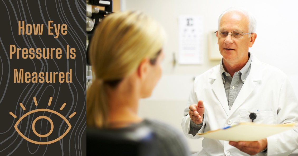
A common question asked during the eye exam is, “When is the puff coming?” Patients are referring to air-puff or non-contact tonometry. Tonometry is the procedure used to measure eye pressure, and this is important for diagnosing and monitoring glaucoma. In non-contact tonometry, a puff of air is used to measure the pressure inside the eye. The benefit of this test is there is no actual contact with the eye, but the air puff is sometimes very startling for patients. Some people hate that test and it isn’t the most accurate way to measure your eye pressure. Some doctors don’t even use the air-puff test. Instead, they place a yellow drop that consists of a numbing medicine and then shine a blue light on the eye. This is done in front of the slit lamp and a small tip gently touches the eye to measure the eye pressure. This procedure is called Goldmann tonometry and is considered the gold standard for measuring eye pressure. Another method for checking eye pressure is the Tonopen. This is a portable, hand-held instrument that is useful when patients can’t sit in front of the slit lamp to have their eye pressure checked. The Tonopen also requires a numbing drop to be placed in the eye, and the tip gently touches the eye. A common question related to tonometry is “what is normal eye pressure?” Normal eye pressure ranges from 10-21 mm Hg. Eye pressure doesn't have any relationship to blood pressure. Many times, people are surprised that their eye pressure is high, but they have normal blood pressure. In general, there is no diet or exercise that will significantly affect eye pressure. It is therefore important to have your eye pressure checked regularly because there are usually no symptoms of high eye pressure until it has affected your vision. Article contributed by Dr. Jane Pan
Diabetic retinopathy is an eye condition that can affect the retina of people who have diabetes. The retina is the light-sensitive tissue that lines the back of the eye, and it detects light that is then processed as an image by the brain. Chronically high blood sugar or large fluctuations in blood sugar can damage the blood vessels in the retina. This can result in bleeding in the retina or leakage of fluid. Diabetic retinopathy can be divided into non-proliferative or proliferative diabetic retinopathy. Non-proliferative diabetic retinopathy: In the early stage of the disease, there is weakening of the blood vessels in the retina that causes out-pouching called microaneurysms. These microaneurysms can leak fluid into the retina. There can also be yellow deposits called hard exudates present in the retina from leaky vessels. Diabetic macula edema is when the fluid leaks into the region of the retina called the macula. The macula is important for sharp, central vision needed for reading and driving. The accumulation of fluid in the macula causes blurry vision. Proliferative diabetic retinopathy: As diabetic retinopathy progresses, new blood vessels grow on the surface of the retina. These blood vessels are fragile, which makes them likely to bleed into the vitreous, which is the clear gel that fills the middle of the eye. Bleeding inside the eye is seen as floaters or spots. Over time, scar tissue can then form on the surface of the retina and contract, leading to a retinal detachment. This is similar to wallpaper contracting and peeling away from the wall. If left untreated, retinal detachment can lead to loss of vision. Symptoms of diabetic retinopathy: Asymptomatic: In the early stages of mild non-proliferative diabetic retinopathy, the person will usually have no visual complaints. Therefore, it is important for people with diabetes to have a comprehensive dilated exam by their eye doctor once a year. Floaters: This is usually from bleeding into the vitreous cavity from proliferative diabetic retinopathy. Blurred vision: This can be the result of fluid leaking into the retina, causing diabetic macular edema. Risk factors for diabetic retinopathy: Blood sugar. Lower blood sugar will delay the onset and slow the progression of diabetic retinopathy. Chronically high blood sugar and the longer the duration of diabetes, the more likely chance of that person having diabetic retinopathy. Medical conditions. People with high blood pressure and high cholesterol are at greater risk for developing diabetic retinopathy. Ethnicity. Hispanics, African Americans, and Native Americans are at greater risk for developing diabetic retinopathy. Pregnancy. Women with diabetes could have an increased risk of developing diabetic retinopathy during pregnancy. If they already have diabetic retinopathy, it might worsen during pregnancy. Article contributed by Jane Pan M.D.

Ceres Eye Care

Built on the foundation of patient convenience and satisfaction, we serve all of your family’s eye care needs under one roof. We're looking forward to seeing you!

© Ceres Eye Care | 1901 Mitchell Road Suite C | Ceres, California 95307 | (209) 537-8971 | Site Map | Email Us
Text and photos provided are the property of EyeMotion and cannot be duplicated or moved.